English
 norsk
norskBlar i forfatter "Ströhl, Florian"
Viser treff 1-20 av 23
-
Adaptive fluctuation imaging captures rapid subcellular dynamics
(Journal article; Tidsskriftartikkel, 2019-07-22)In this work we have explored the live-cell friendly nanoscopy method Multiple Signal Classification Algorithm (MUSICAL) for multi-colour imaging of various organelles and sub-cellular structures in the cardiomyoblast cell line H2c9. We have tested MUSICAL for fast (up to 230Hz), multi-colour time-lapse sequences of various sub-cellular structures (mitochondria, endoplasmic reticulum, microtubules, ... -
Adaptive fluctuation imaging captures rapid subcellular dynamics
(Journal article; Tidsskriftartikkel; Peer reviewed, 2019-07-22)In this work we have explored the live-cell friendly nanoscopy method Multiple Signal Classification Algorithm (MUSICAL) for multi-colour imaging of various organelles and sub-cellular structures in the cardiomyoblast cell line H2c9. We have tested MUSICAL for fast (up to 230Hz), multi-colour time-lapse sequences of various sub-cellular structures (mitochondria, endoplasmic reticulum, microtubules, ... -
Concepts for structured illumination microscopy with extended axial resolution through mirrored illumination
(Journal article; Tidsskriftartikkel; Peer reviewed, 2020-03-20)Wide-field fluorescence microscopy, while much faster than confocal microscopy, suffers from a lack of optical sectioning and poor axial resolution. 3D structured illumination microscopy (SIM) has been demonstrated to provide optical sectioning and to double the resolution limit both laterally and axially, but even with this the axial resolution is still worse than the lateral resolution of ... -
Fluorescence fluctuation-based super-resolution microscopy using multimodal waveguided illumination
(Journal article; Tidsskriftartikkel; Peer reviewed, 2021-07-19)Photonic chip-based total internal reflection fluorescence microscopy (c-TIRFM) is an emerging technology enabling a large TIRF excitation area decoupled from the detection objective. Additionally, due to the inherent multimodal nature of wide waveguides, it is a convenient platform for introducing temporal fluctuations in the illumination pattern. The fluorescence fluctuation-based nanoscopy technique ... -
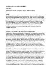
Instant Volume Microscopy of Organoids with SOLIS
(Journal article; Tidsskriftartikkel, 2023)Advancements in microscopy techniques have revolutionized our ability to explore the intricacies of biological systems, with engineered human heart tissue (EHT) being a particularly challenging target. In this article, we will have an in-depth look at the award-winning SOLIS technique (scanned oblique light-sheet instant-volume sectioning), a new twist on multifocus fluorescence microscopy. Through ... -
Label-free nanoscopy enabled by coherent imaging with photonic waveguides
(Journal article; Tidsskriftartikkel; Peer reviewed, 2019-07-29)In this project it was found that Fourier ptychographic microscopy can be improved far beyond its conventional limits via waveguide-based optical systems. Extensive in silico studies showed that images obtained on highrefractive index material waveguide chips in conjunction with hyperspectral illumination light and finely designed waveguide geometries can be combined via a modified phase-retrieval ... -
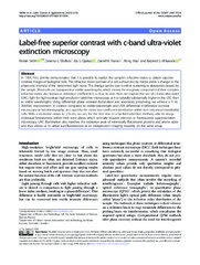
Label-free superior contrast with c-band ultra-violet extinction microscopy
(Journal article; Tidsskriftartikkel; Peer reviewed, 2023-03-03)In 1934, Frits Zernike demonstrated that it is possible to exploit the sample’s refractive index to obtain superior contrast images of biological cells. The refractive index contrast of a cell surrounded by media yields a change in the phase and intensity of the transmitted light wave. This change can be due to either scattering or absorption caused by the sample. Most cells are transparent at visible ... -
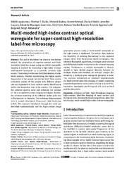
Multi-moded high-index contrast optical waveguide for super-contrast high-resolution label-free microscopy
(Journal article; Tidsskriftartikkel; Peer reviewed, 2022-06-20)The article elucidates the physical mechanism behind the generation of superior-contrast and highresolution label-free images using an optical waveguide. Imaging is realized by employing a high index contrast multi-moded waveguide as a partially coherent light source. The modes provide near-field illumination of unlabeled samples, thereby repositioning the higher spatial frequencies of the ... -
Multifocus microscopy with optical sectioning and high axial resolution
(Journal article; Tidsskriftartikkel; Peer reviewed, 2022)Multifocus microscopy enables recording of entire volumes in a single camera 11 exposure. In dense samples, multifocus microscopy is severely hampered by background haze. 12 Here, we introduce a scalable multifocus method that incorporates optical sectioning and offers 13 improved axial resolution capabilities. In our method, a dithered oblique light-sheet scans the 14 sample volume during a ... -
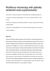
Multifocus microscopy with optically sectioned axial superresolution
(Journal article; Tidsskriftartikkel, 2022)Multifocus microscopy enables recording of entire volumes in a single camera exposure. In dense samples, multifocus microscopy is severely hampered by background haze. Here, we introduce a scalable multifocus method that incorporates optical sectioning and offers axial superresolution capabilities. In our method, a dithered oblique light-sheet scans the sample volume during a single exposure, while ... -
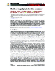
MusiJ: an ImageJ plugin for video nanoscopy
(Journal article; Tidsskriftartikkel; Peer reviewed, 2020-04-14)We present an open-source implementation of the fluctuation-based nanoscopy method MUSICAL for ImageJ. This implementation improves the algorithm’s computational efficiency and takes advantage of multi-threading to provide orders of magnitude faster reconstructions than the original MATLAB implementation. In addition, the plugin is capable of generating super-resolution videos from large stacks of ... -
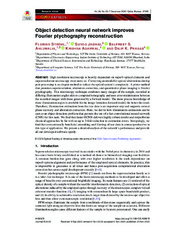
Object detection neural network improves Fourier ptychography reconstruction
(Journal article; Tidsskriftartikkel; Peer reviewed, 2020-11-23)High resolution microscopy is heavily dependent on superb optical elements and superresolution microscopy even more so. Correcting unavoidable optical aberrations during post-processing is an elegant method to reduce the optical system’s complexity. A prime method that promises superresolution, aberration correction, and quantitative phase imaging is Fourier ptychography. This microscopy technique ... -
On-Site Ribosome Remodeling by Locally Synthesized Ribosomal Proteins in Axons
(Journal article; Tidsskriftartikkel; Peer reviewed, 2019-12-10)Ribosome assembly occurs mainly in the nucleolus, yet recent studies have revealed robust enrichment and translation of mRNAs encoding many ribosomal proteins (RPs) in axons, far away from neuronal cell bodies. Here, we report a physical and functional interaction between locally synthesized RPs and ribosomes in the axon. We show that axonal RP translation is regulated through a sequence motif, ... -
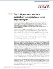
OptiJ: Open-source optical projection tomography of large organ samples
(Journal article; Tidsskriftartikkel; Peer reviewed, 2019-10-30)The three-dimensional imaging of mesoscopic samples with Optical Projection Tomography (OPT) has become a powerful tool for biomedical phenotyping studies. OPT uses visible light to visualize the 3D morphology of large transparent samples. To enable a wider application of OPT, we present OptiJ, a low-cost, fully open-source OPT system capable of imaging large transparent specimens up to 13 mm tall ... -
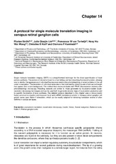
A Protocol for Single-Molecule Translation Imaging in Xenopus Retinal Ganglion Cells
(Chapter; Bokkapittel, 2020-04-29)Single-molecule translation imaging (SMTI) is a straightforward technique for the direct quantification of local protein synthesis. The protein of interest is fused to a fast-folding and fast-bleaching fluorescent protein, allowing one to monitor the appearance of individual fluorescence events after photobleaching of pre-existing proteins in the cell under investigation. The translation of individual ... -
Quantification of the NA dependent change of shape in the image formation of a z-polarised fluorescent molecule using vectorial diffraction simulations
(Journal article; Tidsskriftartikkel; Peer reviewed, 2022-01-19)The point spread function of a fixed fluorophore with its dipole axis colinear to the optical axis appears donut-shaped when seen through a microscope, and its light distribution in the pupil plane is radially polarized. Yet other techniques, such as photolithography, report that this same light distribution in the pupil plane appears as a solid spot. How can this same distribution lead to a spot ... -
Rapid prototyping of 1xN multifocus gratings via additive direct laser writing
(Journal article; Tidsskriftartikkel; Peer reviewed, 2023-04-05)Multifocus gratings (MFGs) enable microscopes and other imaging systems to record entire Z-stacks of images in a single camera exposure. The exact grating shape depends on microscope parameters like wavelength and magnification and defines the multiplexing onto a grid of MxN Z-slices. To facilitate the swift production and alteration of MFGs for a system and application at hand, we have developed ... -
Super-condenser enables labelfree nanoscopy
(Journal article; Tidsskriftartikkel; Peer reviewed, 2019-08-22)Labelfree nanoscopy encompasses optical imaging with resolution in the 100 nm range using visible wavelengths. Here, we present a labelfree nanoscopy method that combines coherent imaging techniques with waveguide microscopy to realize a <i>super-condenser</i> featuring maximally inclined coherent darkfield illumination with artificially stretched wave vectors due to large refractive indices of the ... -
Superresolving the kidney – a practical comparison of fluorescence nanoscopy of the glomerular filtration barrier
(Journal article; Tidsskriftartikkel; Peer reviewed, 2020-12-05)Immunofluorescence microscopy is routinely used in the diagnosis of and research on renal impairments. However, this highly specific technique is restricted in its maximum resolution to about 250 nm in the lateral and 700 nm in the axial directions and thus not sufficient to investigate the fine subcellular structure of the kidney’s glomerular filtration barrier. In contrast, electron microscopy ...